

All Products
-
Nuclear Radiation Protection
-
Nuclear Radiation Detector
-
Copper Foil Shielding
-
RF Shielded Doors
-
RF Shielded Windows
-
Radiation Protection Lead Glass
-
Non Magnetic Tool Kit
-
RF Shielded Chamber
-
Honeycomb Waveguide Air Vents
-
Conductive Adhesive Copper Tape
-
Copper Wire Mesh
-
X Ray Lead Glass
-
EMI Shielding Gasket
-
Electrically Conductive Fabric
-
Radiation Protection Door
-
Radiation Protection X Ray
-
Faraday Cage MRI
-
Copper Wire Wool
-
MRI LED Lighting
-
Non Magnetic Wheelchair
-
Non Magnetic Stretcher
-
 AnasBrass honeycomb vent Looks Very Nice
 -
 SatheeshMRI/RF doors are shining with handles, Thank you My friend.
Non-Magnetic Aluminum Alloy MRI Stretcher with Removable Head Section and 4 Swivel Wheels With Brakes


| Brand Name | Jovi |
|---|---|
| Certification | ISO9001 |
| Model Number | 1-cff-09 |
| Document | JVVM-NMT-Series SPE and Quo...on.pdf |
| Minimum Order Quantity | 1set |
| Price | $1200-$1580 |
| Packaging Details | In Wooden Case Or Customized |
| Delivery Time | 5-8 Work Days |
| Payment Terms | T/T |
| Supply Ability | 300 Set / Month |
Product Details
| Net Weight | 12 Kg | Deck Length | 63 Inches |
|---|---|---|---|
| Head Section Size | 15 X 24 Inches | Total Length | 78 Inches |
| Deck Width | 24 Inches | Height Adjustment | 22 To 32 Inches |
| Highlight | Wheeled Stretcher,Swivel Aluminum,Brake Lock Wheeled Swivel |
||
Product Description
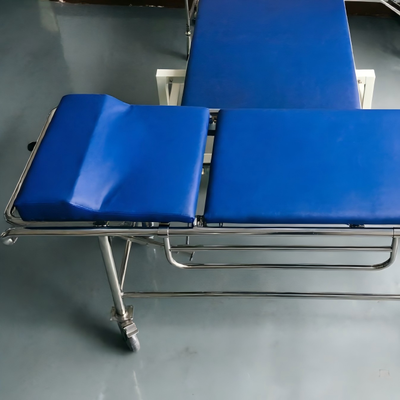
MRI Stretcher with Removable Head Section
The MRI Stretcher with Removable Head Section addresses a specific clinical need: maintaining airway access during MRI scanning. For patients who are intubated, have cervical spine injuries, or are at high risk of respiratory compromise, being able to reach the patient’s airway without moving the entire stretcher is essential. This stretcher solves that problem with a head section that detaches completely in **under 10 seconds**, using no tools and no ferromagnetic components.
The head section measures **15 inches long and 24 inches wide** – the same width as the main deck. When attached, it forms a continuous surface with the rest of the stretcher. The attachment mechanism uses **four locking pins** made of **solid brass** with captured plastic collars. The pins are spring-loaded (using non-magnetic springs) and retract when the release lever is pulled. To detach the head section, the operator pulls two release levers (one on each side) and lifts the section away. Reattachment is equally simple: the section is placed into position, and the pins snap into place automatically.
Once removed, the head section can be placed on a nearby table or held by an assistant. The exposed end of the main deck is rounded and padded, so the patient is not at risk of injury from a sharp edge. The remaining deck length is **63 inches** – still sufficient for patients up to **5 feet 5 inches tall**. For taller patients, the head section can be repositioned rather than removed; it attaches at **three possible positions** (**0, 6, and 12 inches** from the head end), allowing the patient’s head to be positioned at different distances from the scanner bore.
The head section itself is adjustable in angle. It can be set to **0 degrees (flat), 15 degrees of elevation, or 30 degrees of elevation**. The adjustment mechanism uses a non-magnetic gas spring and a plastic ratchet. The gas spring is sealed and requires no maintenance. The angle adjustment is performed by lifting the head section slightly while pressing a release button; the section then raises or lowers under controlled gas spring force.
The removable head section is radiolucent, just like the main deck. This allows portable X-ray of the cervical spine or head without moving the patient, even after the section has been repositioned or removed. The radiolucent property also means that if the patient is being scanned in a head-first position, the head section will not interfere with image quality.
The main deck and head section together provide a total length of **78 inches**. The deck is **24 inches wide**, with a non-slip textured surface. The deck height is adjustable from **22 to 32 inches** using non-magnetic hydraulics. **Four 5-inch casters** with conductive rubber treads and ceramic bearings provide mobility. **Two casters have total-lock brakes; two have directional locks**. The brakes are engaged by foot pedals that provide audible and tactile feedback.
Clinical applications are particularly relevant for anesthesia and critical care. Intubated patients undergoing MRI often require suctioning, ventilator adjustments, or emergency reintubation. With a standard stretcher, accessing the airway may require pulling the patient partially out of the bore – a time-consuming process that interrupts the scan. With this stretcher, the head section can be removed in seconds, providing unrestricted access to the airway while the body remains in position. This feature has been credited with saving at least one life in a reported case where a patient’s endotracheal tube became dislodged during scanning.
The removable head section is also useful for cervical spine imaging. Removing the head section eliminates any potential interference from the stretcher deck, allowing the MRI table’s dedicated head and neck coil to be positioned directly under the patient’s head. The patient’s shoulders remain supported by the main deck, providing stability while the head is fully accessible for coil placement.
Optional accessories include a head holder that attaches to the main deck when the head section is removed, providing lateral support for the patient’s head during scanning. A cervical collar adapter is also available, allowing standard non-magnetic cervical collars to be secured to the stretcher. The stretcher comes with a storage bag for the head section, allowing it to be kept clean and protected when not in use.
Recommended Products